Laboratory Waste
Published:
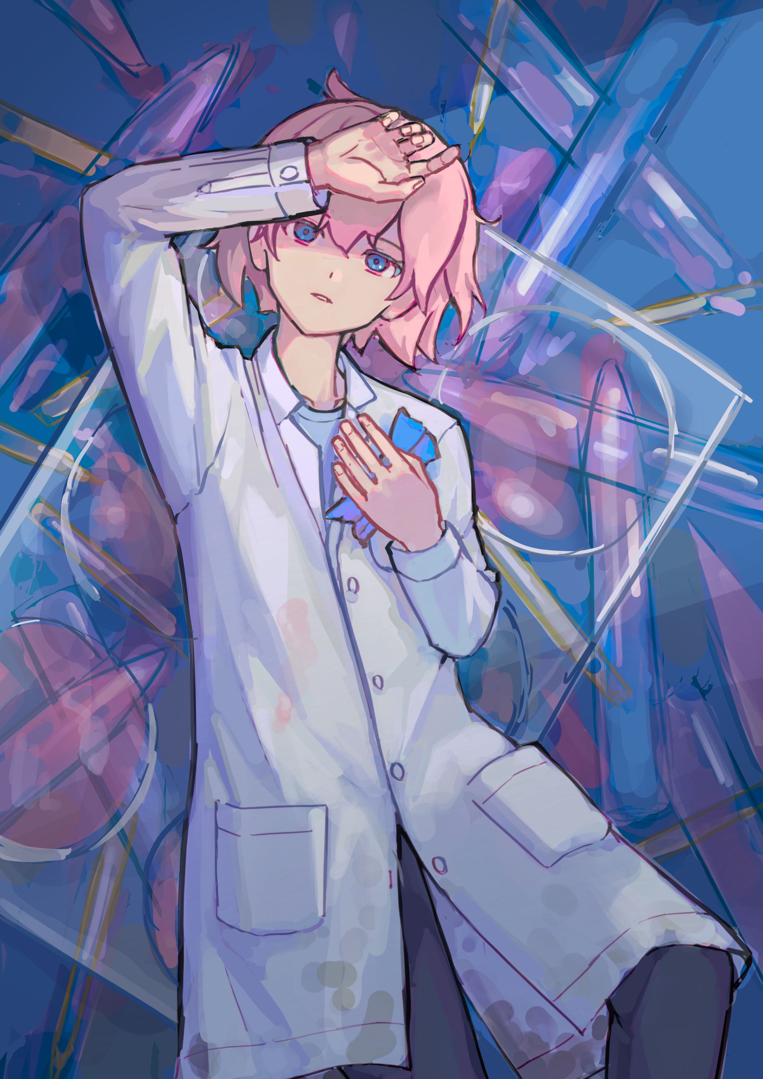
Control the visual hierarchy by applying contrast of hue, contrast of value, and contrast of saturation to illustrate the messy cell culture waste from biomedical laboratories.
Process:
- Rough
- The background is relatively cluttered, so the contrast and overall visual center needs to be controlled. Therefore, the visual of the finished artwork was first considered at the draft stage.
- Contrast of hue: The visual center is a pink color and the background is a blue color, with a translucent pink cell culture medium as a transition between the visual center and the background. The blue latex gloves are placed on the pink area to create a strong contrast.
- Contrast of value: the brightness of the visual center is higher, the brightness of the background is lower.
- Contrast of saturation: the area near the visual center is more saturated, and the area far away from the visual center is less saturated.
- Visual hierarchy guidance: Several pipette tips in the background point to the visual center (character) to guide the viewer’s line of sight.
Lineart
- Adjustments to the structures which were not considered in the rough sketch.
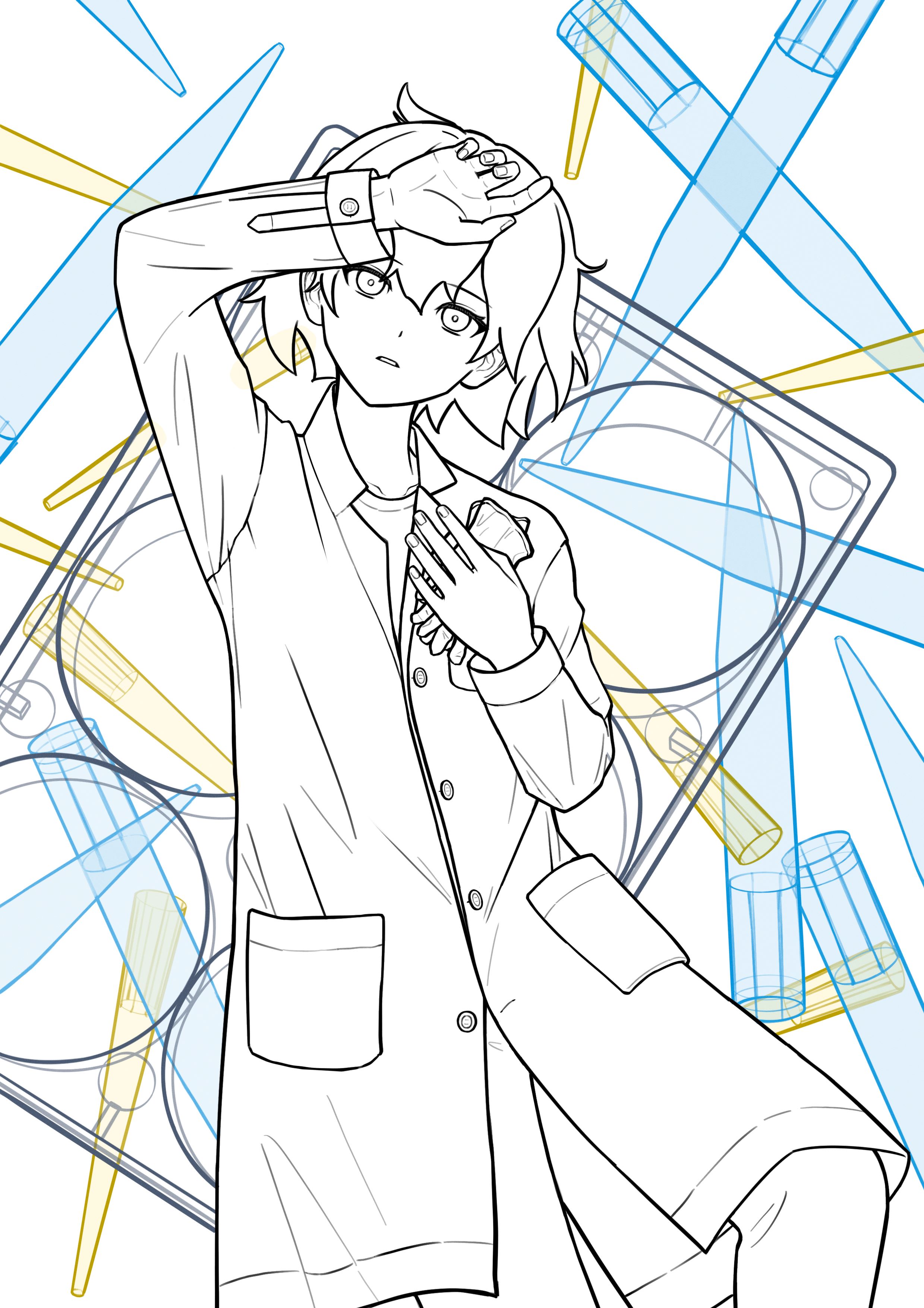
- Adjustments to the structures which were not considered in the rough sketch.
- Rendering to the character
- Changes were made to the color based on the sketch to match the structure of the lineart.
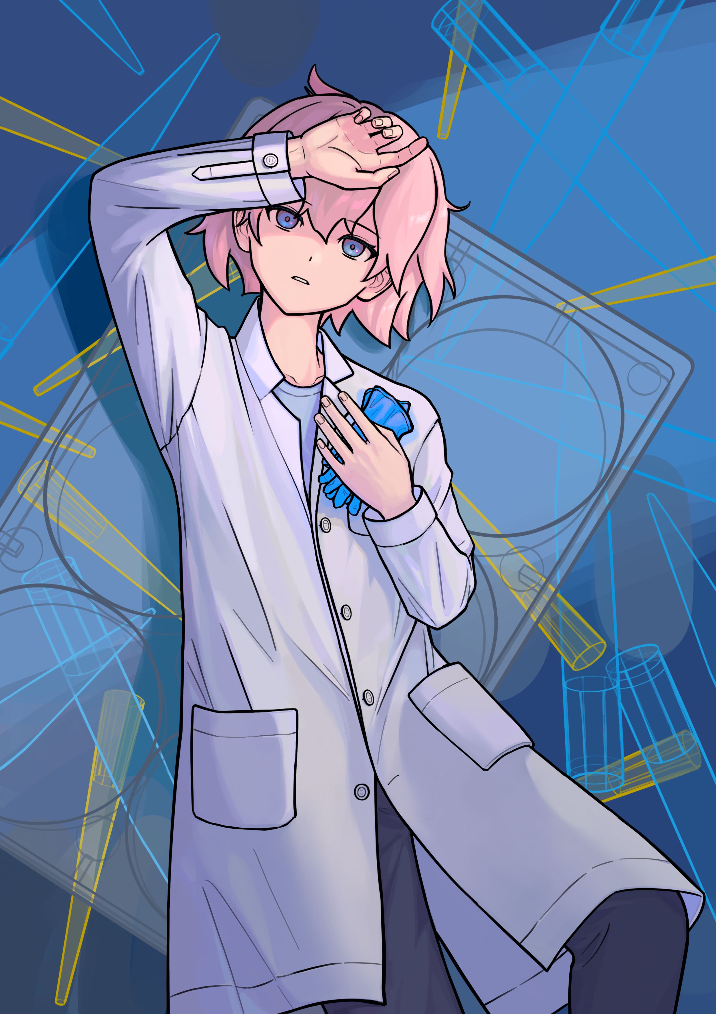
- Changes were made to the color based on the sketch to match the structure of the lineart.
Rendering the background and coloring the lineart

Rendering the 6-well plate

- Rendering the pipette tips
- Also tracing the 6-well plate and the pipette tips that are not covered by it.

- Also tracing the 6-well plate and the pipette tips that are not covered by it.
Adding filters to further reduce the contrast and saturation of the background.

- Update 2025-04-06: Updated character appearance and adjusted the overall colors.

References:
- Pipette tips (200 µL and 1000 µL): Pipette Tips GILSON-fitted (10 / 200 / 1000μl)
- Cell culture medium: Gibco™ Opti-MEM™ I Reduced Serum Medium
- 6-well plate: 6 Well Cell Culture Plate, Flat Bottom, TC Treated, sterile
- Blood stain: Figure 3. a. Less intense ring phenomena were observed on denim textile. b. Positive ring phenomenon observed on bed sheet textile after blood efflux from a neck wound. Adapted from “The ring phenomenon of diluted blood droplets” by F. Ramsthaler et al., 2015, International Journal of Legal Medicine, 130, p.733. Copyright 2015 by Springer-Verlag Berlin Heidelberg

